

Technologies
MRI 1.5 T SIEMENS AVANTO TIM+DOT
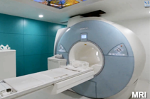
The imaging Centre has State of the art, high-end 1.5 Tesla magnetic resonance imaging scanners that offer increased speed, exceptional resolution, and accuracy, allowing for non-invasive diagnosis of a wide range of conditions. Magnetic resonance imaging (MRI) uses high power magnets and radiofrequency waves instead of x-rays to capture images that give physicians a literal view inside the body. MRI produces soft-tissue images and is used to distinguish normal healthy soft tissue from abnormal diseased tissue. In some instances, a injection of contrast dye may be required.
MRI technology is useful in diagnosing such things
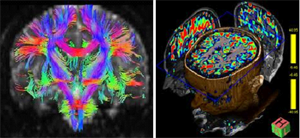
MRI is the ultimate tool for diagnostic imaging and neuroscience research. Providing morphological images with the highest spatial resolution and unmatched soft tissue contrast as well as unique functional information of the CNS in vivo, MRI is the imaging platform for understanding our most complex organ, the brain as infections ,white matter diseases, tumors of brain and brain, strokes at their earliest stages with PERFUSION imaging, neurosurgical planning with TRACTOGRPHY.
It is also used to visualize conditions related to sports injuries, and helps physicians evaluate assess in the soft tissues of the body, bone tumors, cysts, and bulging or herniated discs in the spine. A specialized three-dimensional (3-D) workstation equips specialists with the ability to manipulate
128 SLICE MDCT SIEMENS DEFINATION AS +
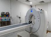
The Imaging Centre is equipped with the 128 slice CT scanner, the latest innovation in CT technology. This state-of-the-art scanner delivers images with increased accuracy and at a faster rate than any other CT scanner available. It also provides three-dimensional (3-D) views, including 3-D views of blood vessels .High speed 128 slice MDCT is very useful in coronary artery imaging, brain perfusion imaging, body imaging and to evaluate bony pathologies.
Computed tomography (CT), often called "CAT" scan, is a system that uses special x-ray equipment to obtain image data and then uses computer processing to show a cross-section of body tissues and organs. Because CT is capable of providing detailed, cross-sectional views of all types of tissue, it is an invaluable tool in studying the chest and abdomen, and is often the preferred method for diagnosing many different cancers. CT examinations are also used to plan and administer radiation treatments for tumors and as a tool to guide physicians performing biopsies or minimally invasive procedures.
CT and MRI images, providing three-dimensional views for the most comprehensive study and accu-Rate diagnosis. 3dimensional images of the inside of the human body, helping in the treatment of conditions such as cancer and heart disease.
High end Ultrasound and Doppler machine GE LOGIQ S7
The Imaging Centre is equipped with the High end Ultrasound and Doppler machines for sonographic needs, including abdominal, musculoskeletal imaging, breast, pelvic, and gynecological exams.
SIEMENS 3000 NOVA Mammography machine
The Imaging Centre is equipped with high-end, state of the art Mammography machine located in the same building. The Breast Care Centre is equipped with digital mammography machine and a specialized USG machine with Matrix probe. Breast imaging specialists use the latest technology to perform minimally invasive image-guided breast biopsies.
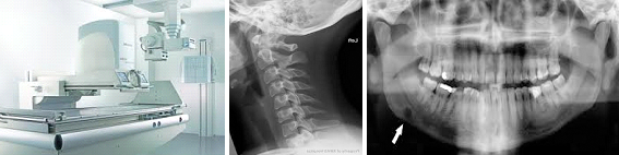
500 Ma X RAY MACHINE WITH DIGITAL (CR)RADIOGRAPHY
The Imaging Centre is equipped with the Digital (CR) Radiography, hig-hend X-RAY machine and, the latest OPG machine. This state-of-the-art machines delivers images with increased accuracy and at a faster rate.